
Fig. 2
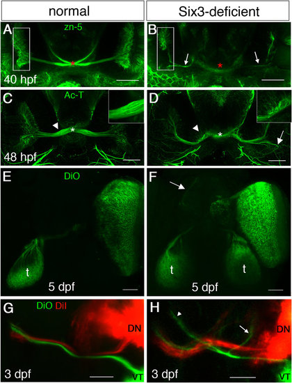
Optic nerve misrouting in Six3-deficient embryos. (A-H) Maximum projection confocal images of labelled RGC axons in WT (A,C,E,G) and Six3-deficient (B,D,F,H) embryos. (A,B) zn-5 immunohistochemistry at 40 hpf. Asterisk in A marks the optic chiasm and in (B) where the optic chiasm should be. Arrows in B point at RGC axons. Rectangles in (A,B) mark the GCL of one eye. (C,D) Ac-T immunohistochemistry at 48 hpf. Asterisks in (C,D) mark location of the optic chiasm. Arrow in (D) points at a region where RGC axons are misrouted and fail to exit the eye. Insets in (C,D) show higher magnification of the regions at which arrowheads point. (E,F) DiO anterograde labelling from a single eye at 5 dpf. Arrow in F points at axons reaching the forebrain. (G,H) DiO (green) and DiI (red) labelling of VT and ND RGCs, respectively, at 3 dpf. t, tectum. In (H) arrow points at VT axons remaining ipsilaterally and arrowhead points at VT axons misrouted to the forebrain. (A-D) are ventral views and E-H are dorsal views. Anterior is up. Scale bars are 50 µm. |
| Antibodies: | |
|---|---|
| Fish: | |
| Anatomical Terms: | |
| Stage Range: | Prim-25 to Long-pec |
| Fish: | |
|---|---|
| Observed In: | |
| Stage Range: | Prim-25 to Day 5 |