
FIGURE
Fig. S11
- ID
- ZDB-FIG-160303-33
- Publication
- Chen et al., 2016 - Efficient extravasation of tumor-repopulating cells depends on cell deformability
- Other Figures
- All Figure Page
- Back to All Figure Page
Fig. S11
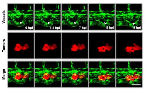
High resolution microscopy of time-course of TRCs extravasation. TRCs arrested at the blood vessels of the zebrafish tail extravasated gradually into peripheral tissues. Time-lapse images were acquired using confocal microscopy at 30-min intervals. White arrowheads indicated the boundaries of vessels. White dashed lines circled projected areas of extravasated TRCs. Color code: zebrafish blood vessels are green, and melanoma cells are red. Scale bar: 50 µm. |
Expression Data
Expression Detail
Antibody Labeling
Phenotype Data
Phenotype Detail
Acknowledgments
This image is the copyrighted work of the attributed author or publisher, and
ZFIN has permission only to display this image to its users.
Additional permissions should be obtained from the applicable author or publisher of the image.
Full text @ Sci. Rep.