
Fig. 7
- ID
- ZDB-FIG-160303-25
- Publication
- Chen et al., 2016 - Efficient extravasation of tumor-repopulating cells depends on cell deformability
- Other Figures
- All Figure Page
- Back to All Figure Page
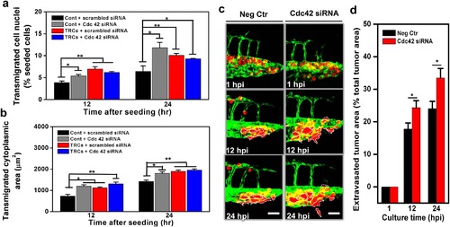
Silencing Cdc42 in control melanoma cells elevates transmigration in vitro and extravasation in vivo.Transmigration of control melanoma cells or of TRCs through 3-µm pore membrane transwell was measured after the cells were transfected with siRNA to Cdc42 or scrambled siRNA. (a) Transmigrated cell nuclei is the percentage of transmigrated cell nuclei (% seeded cells) per view-field. (b) Transmigrated cytoplasmic area is the total transmigrated cytoplasmic area per view-field. Mean + s.e.m.; n = 3 independent experiments. *p < 0.05, **p < 0.01, ***p < 0.001. (c) Control melanoma cells transfected with scrambled siRNA (Neg Ctr) or Cdc42 siRNA were injected into the pericardium of 48 hpf embryos respectively. Images in each panel show vessel penetration of Neg Ctr (left panels) or Cdc42 siRNA (right panels) at 1, 12, and 24 hpi respectively. Dashed white lines mark the tumor extravasation areas (i.e., various sizes of micrometastases) from vessels to surrounding tissues. Scale bars, 50 µm. (d) Quantification of extravasated tumor area relative to the total tumor area at different time points: 1, 12, and 24 hpi. Color code: Zebrafish blood vessels are green, and mouse tumor cells are red. Mean + s.e.m.; n >10 fish per group from 3 independent experiments; *p < 0.05. |