
Fig. S1
- ID
- ZDB-FIG-160303-27
- Publication
- Chen et al., 2016 - Efficient extravasation of tumor-repopulating cells depends on cell deformability
- Other Figures
- All Figure Page
- Back to All Figure Page
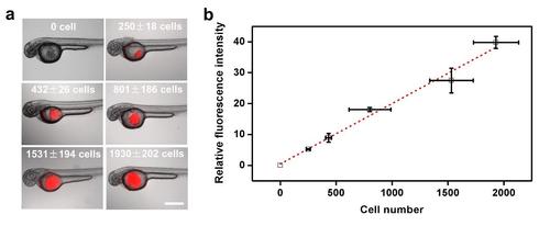
Calibration of cell number in zebrafish. (a) Representative images of 2 dpf zebrafish injected with different numbers of tumor cells into the yolk. Red color are tfRFP B16 melanoma cells. (b) Calibration of the number of injected tumor cells into the yolk with relative fluorescence intensity (corresponding to the projected fluorescence area multiplying fluorescence intensity). Injected cells were counted using a cell counter in vitro before and after injection. Projected fluorescence area and intensity were quantified using ImageJ. Dashed line is the linear fit of the data. Mean±s.e.m.; n≥10 larvae. Scale bar, 500 µm. |