
Fig. 2
- ID
- ZDB-FIG-160303-22
- Publication
- Chen et al., 2016 - Efficient extravasation of tumor-repopulating cells depends on cell deformability
- Other Figures
- All Figure Page
- Back to All Figure Page
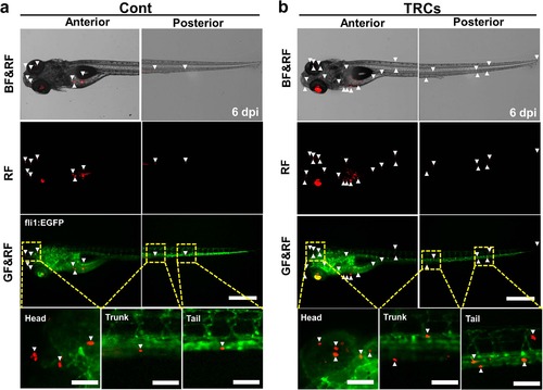
TRCs exhibit more metastases.The same number (~500 cells) of tfRFP melanoma cells cultured on 2D rigid plastic for 5 days (Cont) or in 3D 90-Pa fibrin gels for 5 days (TRCs) was injected into the yolk of 2 dpf embryos respectively. Representative images show metastatic tumor foci at 6 dpi zebrafish, comparing the TRCs group (b) with the Cont group (a). Arrowheads point to disseminated tumor foci (single tumor cells or tumor cell aggregates). There are more arrowheads in the TRCs group, suggesting more metastases. Color code: Zebrafish blood vessels are green, and melanoma cells are red; BF = brightfield; GF = Green Fluorescence; RF = tfRFP Fluorescence. Scale bars, 500 µm. |