
Fig. S9
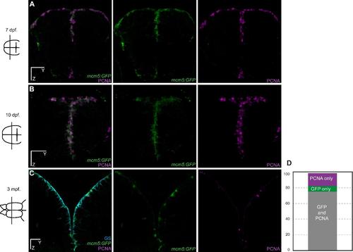
Validation of the Tg(mcm5:eGFP)gy2 transgenic line for the reliable reporting of neural progenitor cell division in the zebrafish pallium. (A-C) Cross-sections of the telencephalon are shown in transgenic zebrafish at 7 day-postfertilization (dpf), 10 dpf and 3 month-post-fertilization (mpf) with double immunostaining for the proliferation marker PCNA (magenta) and GFP (green). (D) Quantification showing that GFP is strictly co-expressed with PCNA in 75% (sem=3.1) of the positive ventricular progenitors (sum of PCNA and MCM5 cells). 17% (sem=4.0) of the positive cells express GFP only, which we interpret as cells having divided but still maintaining some GFP protein due to GFP stability. 8% (sem=1.8) of the positive cells express PCNA only, which we interpret as cell just entering the cell cycle with no GFP detectable yet. n = 4 brains each with 3 to 6 sections per brain. Scale bars: 30 µm (A-C). |