
FIGURE
Fig. 3
- ID
- ZDB-FIG-151109-5
- Publication
- Turola et al., 2015 - Ovarian senescence increases liver fibrosis in humans and zebrafish with steatosis
- Other Figures
- All Figure Page
- Back to All Figure Page
Fig. 3
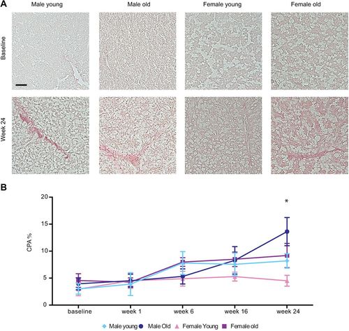
Assessment of hepatic fibrosis in overfed zebrafish. (A) Sirius Red staining of liver sections from young males, old males, young females and old female zebrafish at baseline, and after 24weeks of overfeeding. Scale bar: 30µm. (B) Quantification of liver fibrosis by using computer-assisted digital image analysis. Data are reported as collagen proportionate area (CPA) converted into a percentage. Three sections from five fish per subgroup were analyzed. Values are expressed as means±s.d. *P<0.0001 for young females versus all the other groups. |
Expression Data
Expression Detail
Antibody Labeling
Phenotype Data
Phenotype Detail
Acknowledgments
This image is the copyrighted work of the attributed author or publisher, and
ZFIN has permission only to display this image to its users.
Additional permissions should be obtained from the applicable author or publisher of the image.
Full text @ Dis. Model. Mech.