
FIGURE
Fig. S1
- ID
- ZDB-FIG-151109-6
- Publication
- Turola et al., 2015 - Ovarian senescence increases liver fibrosis in humans and zebrafish with steatosis
- Other Figures
- All Figure Page
- Back to All Figure Page
Fig. S1
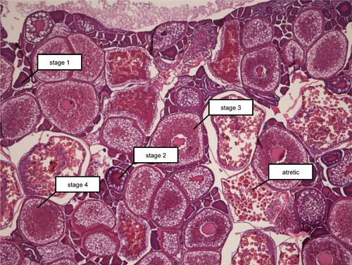
Developmental stages of the oocytes. Stage 1, perinucleolar oocytes. The oocytes size was small and ooplasm lacks granular structure. Stage 2, cortical alveolar oocytes. The vitelline envelope (zona radiata) begin to form. Stage 3, vitellogenic oocyte. The oocyte increased in size; the granular structures in ooplasm were larger and the nucleus was irregular in the shape. Stage 4, mature oocyte. The nucleus could not be observed due to the granular structures that filled up the entire cytoplas. Atretic stage. Oocyte and the vitelline membrane structure started to disintegrate. |
Expression Data
Expression Detail
Antibody Labeling
Phenotype Data
Phenotype Detail
Acknowledgments
This image is the copyrighted work of the attributed author or publisher, and
ZFIN has permission only to display this image to its users.
Additional permissions should be obtained from the applicable author or publisher of the image.
Full text @ Dis. Model. Mech.