
FIGURE
Fig. S2
Fig. S2
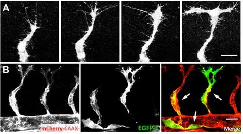
(A) Confocal image time series of a single growing trunk ISV sprout in a Tg(fli1a:EGFP)y1 embryo, showing dynamic changes in sprout morphology over time. (B) Confocal image of growing trunk ISV in a 28 hpf Tg(kdrl:mCherry-caax)y171 embryo injected with a Tol2(fli1a:EGFP-F)y288 transgene. The injected transgene mosaically marks subsets of endothelial cells, but it is not possible to determine whether contiguously labeled endothelium represents one cell or multiple adjacent cells. All images are lateral views with rostral to the left. Scale bars = 20 µm. |
Expression Data
Expression Detail
Antibody Labeling
Phenotype Data
Phenotype Detail
Acknowledgments
This image is the copyrighted work of the attributed author or publisher, and
ZFIN has permission only to display this image to its users.
Additional permissions should be obtained from the applicable author or publisher of the image.
Full text @ Development