
Fig. 3
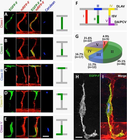
Heterogeneous morphology of individual ECs in newly forming ISV/DLAV segments. (A-E) Representative confocal micrographs of verified single Tol2(fli1a:H2B-TagBFP-p2A-egfp-F) transgene-expressing ECs in trunk ISV/DLAV segments in a 42hpf Tg(kdrl:mRFP-F)y286 germline transgenic embryo, showing green, red, red/green merge and blue fluorescent channels. (F) Morphological classification of ECs contributing to the ISV/DLAV. Class I, extending along an entire ascending ISV segment (see A); Class II, extending partially along an entire ascending ISV (see B); Class III, exclusively in the DLAV (see C); Class IV, in both DLAV and ISV (see D); Class V, in both DA and ISV (see E). (G) Quantification of the proportion of ECs found in each of the five morphological classes. (H,I) High-magnification GFP fluorescence (H) and merged GFP/BFP/RFP fluorescence (I) images of a representative verified single Tol2(fli1a:H2B-TagBFP-p2A-egfp-F) transgene-expressing EC in a trunk ISV segment in a 42hpf Tg(kdrl:mRFP-F)y286 germline transgenic embryo, showing the morphological complexity of the cell, with fine processes extending up and down the vessel segment past the main part of the EC body. PCV, posterior cardinal vein. Scale bars: 20µm in A-E; 10µm in H,I. |