
FIGURE
Fig. 4
- ID
- ZDB-FIG-150824-8
- Publication
- Crucke et al., 2015 - The innervation of the zebrafish pharyngeal jaws and teeth
- Other Figures
- All Figure Page
- Back to All Figure Page
Fig. 4
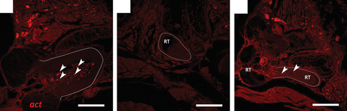
Immunohistochemical detection of nerve fibres in both functional teeth (full line) and replacement teeth (dashed line), on transverse paraffin sections of adult zebrafish stained with a primary antibody against acetylated tubulin (act). (A) Functional teeth clearly contain many small nerve fibres (arrowheads) in the dental pulp. (B) Replacement teeth (RT), however, are completely devoid of axons at all stages of differentiation. (C) Only at stages of very late cytodifferentiation, i.e. nearing attachment, could nerves (arrowheads) be seen entering the tooth at the base. Scale bars: 50 µm (A); 150 µm (B, C). |
Expression Data
Expression Detail
Antibody Labeling
Phenotype Data
Phenotype Detail
Acknowledgments
This image is the copyrighted work of the attributed author or publisher, and
ZFIN has permission only to display this image to its users.
Additional permissions should be obtained from the applicable author or publisher of the image.
Full text @ J. Anat.