
Fig. 1
- ID
- ZDB-FIG-150824-5
- Publication
- Crucke et al., 2015 - The innervation of the zebrafish pharyngeal jaws and teeth
- Other Figures
- All Figure Page
- Back to All Figure Page
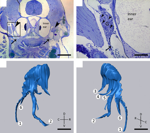
(A, B) Toluidine blue-stained transverse sections of 8.3 mm SL zebrafish showing the presence of two large ganglia (G) (A, arrowheads) dorsal to the developing dentition, lateral to the inner ear, and apposed to the posterior cardinal vein (arrow, A, B). Larger magnification of the area indicated in (A) clearly shows the cluster of neuronal cell bodies (B, arrowheads). (C, D) 3D reconstruction of the ganglion shown in (A) on the right body side. Note the six different branches, two of which (1, 2) have a common origin and course ventrally, three branches run medially (3, 4, 5), and a final branch runs caudally (6). C, caudal; D, dorsal; R, rostral; V, ventral. Scale bars: 200 µm (A); 50 µm (B–D). |