
- Title
-
The innervation of the zebrafish pharyngeal jaws and teeth
- Authors
- Crucke, J., Van de Kelft, A., Huysseune, A.
- Source
- Full text @ J. Anat.
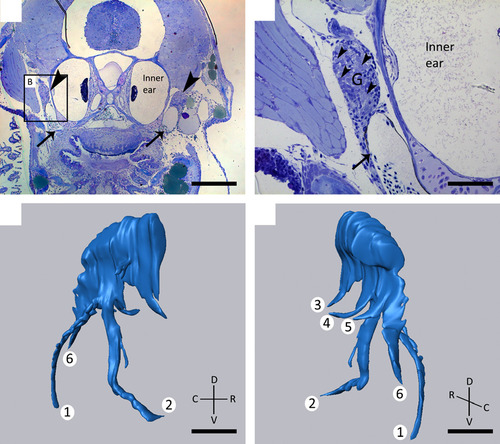
(A, B) Toluidine blue-stained transverse sections of 8.3 mm SL zebrafish showing the presence of two large ganglia (G) (A, arrowheads) dorsal to the developing dentition, lateral to the inner ear, and apposed to the posterior cardinal vein (arrow, A, B). Larger magnification of the area indicated in (A) clearly shows the cluster of neuronal cell bodies (B, arrowheads). (C, D) 3D reconstruction of the ganglion shown in (A) on the right body side. Note the six different branches, two of which (1, 2) have a common origin and course ventrally, three branches run medially (3, 4, 5), and a final branch runs caudally (6). C, caudal; D, dorsal; R, rostral; V, ventral. Scale bars: 200 µm (A); 50 µm (B–D). |
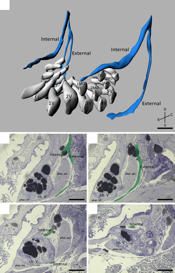
(A) 3D visualization of a 9.5 mm SL zebrafish dentition demonstrating the main nerve branches (blue) in the vicinity of the developing teeth (white). The internal and external branches emerge from a common stem on the postero-dorsal side of the dentition. Note that the internal branch is split up in two bundles on one body side. The teeth are organized in a ventral (1V–5V), mediodorsal (1MD–4MD) and dorsal (1D–2D) row. (B–E) Consecutive sagittal toluidine blue-stained sections of a wild-type zebrafish (SL = 8.3 mm) showing how the internal and external branch (pseudocoloured green) course in relation to both internal and external pharyngoclavicularis muscles (phar. int./ phar. ext.), and the fifth ceratobranchials (cb. V). The external branch extends ventrally and bends in a medial direction along the ventral side of the dentition. The internal branch on the other hand, bends medially almost instantly thus running along the dorsal side of the dorsal tooth row. C, caudal; D, dorsal; R, rostral; V, ventral. Scale bars: 50 µm (A); 100 µm (B–E). |
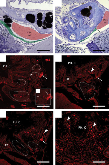
(A, B) Sagittal toluidine blue-stained sections of a wild-type 9.5 mm SL zebrafish. In both sections anterior is to the right. The external branch (pseudocoloured green) passes close to the external pharyngoclavicularis muscle (phar. ext, pseudocoloured red) at the posterior side of the dentition (A). The external branch appears to terminate (arrow) at the internal pharyngoclavicularis (phar. int) muscle on the cranioventral side of the dentition (B).(C–F) Immunohistochemical detection of nerve fibres in the dentition of zebrafish. Transverse paraffin sections of adult fish were stained with a primary antibody against acetylated tubulin (act). Functional teeth are indicated using dashed lines. (C) The internal branch (arrow) passes at the very base of the functional teeth (dashed line) after having penetrated the ceratobranchial bone at tooth position 1d (not shown). (C′) Enlargement of the functional tooth indicated in (c) (*); smaller branches appear to enter the pulp cavity of the tooth. (D, E) Furthermore, smaller axons (arrowheads) extend from the internal branch (arrow) towards the pharyngeal epithelium, where they probably innervate the oral mucosa and taste buds located between the epithelial crypts (F). PH. C., pharyngeal cavity; RT, replacement tooth. Scale bars: 100 µm (A–E); 50 µm (F). |
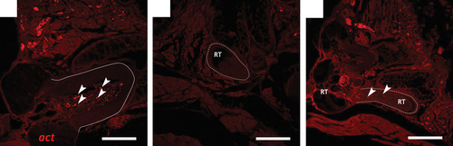
Immunohistochemical detection of nerve fibres in both functional teeth (full line) and replacement teeth (dashed line), on transverse paraffin sections of adult zebrafish stained with a primary antibody against acetylated tubulin (act). (A) Functional teeth clearly contain many small nerve fibres (arrowheads) in the dental pulp. (B) Replacement teeth (RT), however, are completely devoid of axons at all stages of differentiation. (C) Only at stages of very late cytodifferentiation, i.e. nearing attachment, could nerves (arrowheads) be seen entering the tooth at the base. Scale bars: 50 µm (A); 150 µm (B, C). |