
Fig. 3
- ID
- ZDB-FIG-150824-7
- Publication
- Crucke et al., 2015 - The innervation of the zebrafish pharyngeal jaws and teeth
- Other Figures
- All Figure Page
- Back to All Figure Page
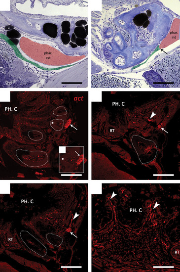
(A, B) Sagittal toluidine blue-stained sections of a wild-type 9.5 mm SL zebrafish. In both sections anterior is to the right. The external branch (pseudocoloured green) passes close to the external pharyngoclavicularis muscle (phar. ext, pseudocoloured red) at the posterior side of the dentition (A). The external branch appears to terminate (arrow) at the internal pharyngoclavicularis (phar. int) muscle on the cranioventral side of the dentition (B).(C–F) Immunohistochemical detection of nerve fibres in the dentition of zebrafish. Transverse paraffin sections of adult fish were stained with a primary antibody against acetylated tubulin (act). Functional teeth are indicated using dashed lines. (C) The internal branch (arrow) passes at the very base of the functional teeth (dashed line) after having penetrated the ceratobranchial bone at tooth position 1d (not shown). (C′) Enlargement of the functional tooth indicated in (c) (*); smaller branches appear to enter the pulp cavity of the tooth. (D, E) Furthermore, smaller axons (arrowheads) extend from the internal branch (arrow) towards the pharyngeal epithelium, where they probably innervate the oral mucosa and taste buds located between the epithelial crypts (F). PH. C., pharyngeal cavity; RT, replacement tooth. Scale bars: 100 µm (A–E); 50 µm (F). |