
Fig. S3
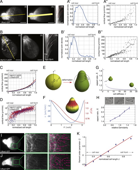
Actomyosin Cortical Flows and Spatial Cortical Density Gradients Determine the Shape of Polarized Stable-Bleb Cells, Related to Figure 4 (A) TIRFM image (I) of a polarized stable-bleb cell obtained from Tg(actβ1:lifeact-GFP) transgenic embryos at sphere stage (4 hpf) and cultured in confinement under agarose on a PEG-coated substrate in the presence of 30 µM LPA. (II) Averaged fluorescence image over 50 frames and (III) kymograph data along yellow line in (I). (A′) Single cell density profile along yellow line in (II). Width of yellow line in (II) indicates the lateral line extent used for calculating average density profiles. (A′′) Single cell cortical flow profile obtained from kymograph data in (III). (B) TIRFM image (I) of a polarized stable-bleb cell obtained from Tg(actβ1:myl12.1eGFP) transgenic embryos at sphere stage (4 hpf) and cultured in confinement under agarose on a PEG-coated substrate in the presence of 30 µM LPA. (II) Average fluorescence image over 50 frames and (III) kymograph along the axis of polarization. (B′ and B′′) Single cell density (B′) and cortical flow profile (B′′) obtained along yellow line in (II) and kymograph data (III). (C) Average cortical flow profile of data for Myl12.1eGFP (gray, mean ± sem; n = 27) with exponential fit (blue) and Lifeact-GFP (black, mean ± sem; n = 30) with exponential fit (red). (D) Log-Linear plot of single cell cortical flow data obtained from (C). (E) Illustration of cell shape change resulting from cortical tension. The un-deformed state of the cell is assumed to be spherical with radius R. An arbitrary point P in the cytoplasm gets shifted to P2 under deformation, by an amount u (vector). (F) Cortical flow and density profile in the polarized state of the cell. In the steady state, inhomogeneous solutions to v(Θ) and ρ(Θ) determine the cortical tensions and friction that are exerted on the bulk of the cell, and, as a result, the cell shape in the polarized state (shown in inset). The parameter values for the determination of v(Θ) and ρ (Θ), and the cell deformation are: ζ β2/ξR2Χ=2, η/ξR2=0.1, (R4ξ/ρ20γ)kd=1, β=1, and (R4ξ/ρ20γ)Χ=1. The cortical flow is indicated in µm/min (taking kd = 0.1 s1 (Pollard and Borisy, 2003) and R = 20 µm), while the actin density is expressed relative to the unperturbed value, ρ0 (which, without loss of generality, we take to be equal to one). (G) Phase diagram of cell shapes showing cell aspect ratio A versus cell stiffness E. (H) Representative bright-field images of polarized progenitor cells cultured at varying osmotic conditions in the presence of 30 µM LPA with numbers indicating relative medium osmolarities (standard medium 1.0, hypoosmotic 0.75, hyperosmotic 1.25; top) and quantification of cell aspect ratios for varying medium osmolarities (n = 50 cells for all conditions). (I and J) TIRF images of Lifeact-GFP localization in polarized progenitor cells obtained from Tg(actβ1:lifeact-GFP) embryos cultured in non-adhesive confinement under agarose in the presence of 30 µM LPA (left). Magnification of green highlighted area (middle) and tracking of filament alignment (right). (K) Quantification of filament alignment order parameter Q along the normalized cell length x with theoretical fit function (see Equations S29, S30, and S31). All scale bars, 10 µm. |
Reprinted from Cell, 160, Ruprecht, V., Wieser, S., Callan-Jones, A., Smutny, M., Morita, H., Sako, K., Barone, V., Ritsch-Marte, M., Sixt, M., Voituriez, R., Heisenberg, C.P., Cortical Contractility Triggers a Stochastic Switch to Fast Amoeboid Cell Motility, 673-685, Copyright (2015) with permission from Elsevier. Full text @ Cell