
Fig. S4
- ID
- ZDB-FIG-150316-16
- Publication
- Maier et al., 2014 - RA and FGF Signalling Are Required in the Zebrafish Otic Vesicle to Pattern and Maintain Ventral Otic Identities
- Other Figures
- All Figure Page
- Back to All Figure Page
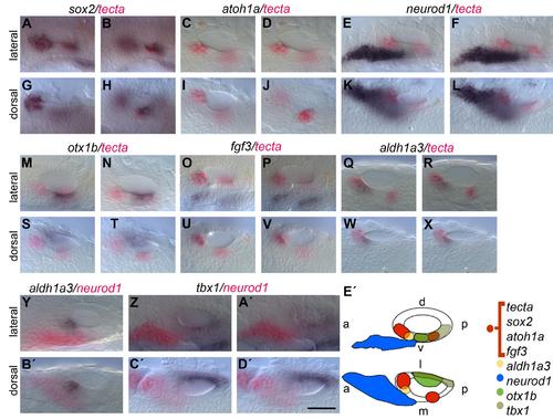
Wild-type expression pattern of sox2, atoh1a, tecta, neurod1, tbx1, otx1b, fgf3 and aldh1a3 in the zebrafish otic vesicle at 26 hpf. (A,B,G,H) sox2 (purple) and tecta (red) are co-expressed in the presumptive anterior and posterior maculae. Expression of sox2 (purple) is broader compared with expression of tecta. (C,D,I,J) atoh1a (weak purple) and tecta (red) are co-expressed in the presumptive anterior and posterior maculae. Expression of atoh1a (purple) is more restricted compared with expression of tecta. (E,F,K,L) The neurogenic marker neurod1 (purple) is mainly expressed in neuroblasts of the statoacoustic ganglion beneath the OV; expression of tecta (red) marks the developing sensory maculae in the otic epithelium. (M,N,S,T) The non-neural marker otx1b (purple) and tecta (red) are expressed in distinct domains in the OV. Expression of otx1b can be detected in a ventrolateral domain. (O,P,U,V) fgf3 (purple) and tecta (red) are co-expressed in the anterior OV. (Q,R,W,X) aldh1a3 (purple) is expressed in the anterior OV, partially overlapping with the expression domain of tecta (red) but extending more ventromedially. (Y,B′) aldh1a3 (purple) is expressed in the anterior OV in a position next to the expression domain of neurod1 (red). (Z,A′,C′,D′) tbx1 (purple) is expressed in the ventrolateral OV, posterior to the expression domain of neurod1 (red). (E′) Schematic representation of the expression domains in relation to each other. a: anterior, p: posterior, d: dorsal, v: ventral, l: lateral, m: medial. Scale bar: 50 µm. |