
Fig. S5
- ID
- ZDB-FIG-141023-39
- Publication
- Pope et al., 2014 - Peripheral glia have a pivotal role in the initial response to axon degeneration of peripheral sensory neurons in zebrafish
- Other Figures
- All Figure Page
- Back to All Figure Page
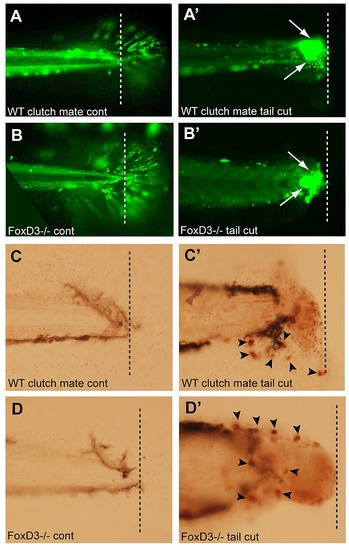
Loss of foxD3 does not result in an impaired immune response. Tail transections were performed on 4 dpf foxD3-/-;mpx:gfp or WT clutch mates. Larvae were anesthetized with tricaine and tails were transected with a scalpel at the junction of the body and tail fin. 2 hr after the tail transection, larvae were either imaged for GFP+ cells (mpx:gfp, neutrophils) or stained with NR to visualize macrophages. A) Uncut tail of WT clutch mate shows the location of the tail transection (dashed line in all images). A′) Transection site of WT clutch mate showed an increase in neutrophils at the location of the cut. B) Uncut tail of foxD3-/-;mpx:gfp shows location of tail transection. B′) Transected tail of foxD3-/-;mpx:gfp also showed an increase in neutrophils at the location of the cut (arrow), similar to that seen in WT clutch mates. C) Neutral Red staining in uncut tail of WT clutch mate. C′) NR staining at transection of WT clutch mate (arrow) showed an increase in macrophages at the location of the cut. D) NR staining at the transection site in a foxD3-/-;mpx:gfp larva. D′) NR staining of transection site in a foxD3-/-;mpx:gfp showed an increase in macrophages at the location of the transection (arrow), similar to that seen in WT clutch mates. |