
Fig. 7
- ID
- ZDB-FIG-141023-34
- Publication
- Pope et al., 2014 - Peripheral glia have a pivotal role in the initial response to axon degeneration of peripheral sensory neurons in zebrafish
- Other Figures
- All Figure Page
- Back to All Figure Page
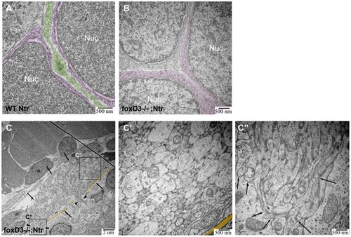
TEM images of missing peripheral glia in FoxD3-/-Ntr. A) Ganglion in 4 dpf wild type Ntr (40,000x) shows two cell bodies (nuclei, labeled Nuc; cytoplasm in magenta) with satellite glia (green) located in between. B) Ganglion in 4 dpf FoxD3-/-Ntr (30,000x) shows three adjacent cell bodies (nuclei in orange; cytoplasm in magenta) without satellite glia present. C,C′,C′′) Axons in 4 dpf FoxD3-/-;Ntr. C) Cross section of axon bundle (arrows) and longitudinal view of axons entering hindbrain (arrowheads) (7500x). Basement membrane indicated in orange in all images. C′) Higher magnification (30,000x) of axon bundle. Medium small axons appear relatively normal (compare to wild-type Ntr in Figure 4C′). C′′) Higher magnification (30,000x) shows defasciculated axons (arrows) entering brain (compare to Figure 4C′′). |
| Fish: | |
|---|---|
| Condition: | |
| Observed In: | |
| Stage: | Day 4 |