
Fig. 4
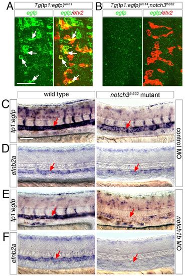
Multiple Notch receptors contribute to artery differentiation. (A,B) Confocal micrographs of flat-mounted sibling embryos at 10 ss following fluorescence in situ hybridization with riboprobes against egfp (green) and etv2 (red). Dorsal view, anterior is up. (A) Wild type Tg(tp1:egfp)um14; arrows indicate etv2/egfp double-positive cells. (B) Tg(tp1:egfp)um14;notch3fh332 mutant sibling. (C-F) DIC images of Tg(tp1:egfp)um14 wild-type (left panels) and notch3fh332 mutant (right panels) embryos at 24 hpf subjected to in situ hybridization with riboprobes against egfp (C,E) or efnb2a (D,F); (C,D) Embryos injected with 2.5 ng control morpholino (MO). (E,F) Embryos injected with 2.5 ng notch1b MO. Scale bars: 40 μm (A,B); 50 μm (C-F). |
| Genes: | |
|---|---|
| Fish: | |
| Knockdown Reagent: | |
| Anatomical Terms: | |
| Stage Range: | 10-13 somites to Prim-5 |