
Fig. 8
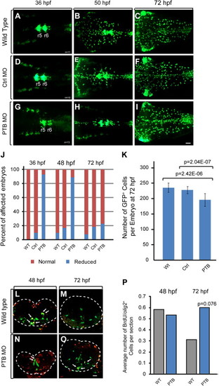
Increased replication of OPCs in prdm12b deficient embryos. (A–I) olig2:egfp Expression in wild type (A–C), control MO-injected (D–F), or PTB MO-injected (G–I) embryos. (J) Quantification of embryos with reduced numbers of olig2:egfp+ cells. (K) Quantification of olig2:egfp+ cells per embryo at 72 hp. (L–O) Cross sections of wild type (L, M) and PTB MO-injected (N, O) Tg(olig2:egfp) embryos labeled with anti-BrdU. (P) Quantification of the number of olig2:egfp+/BrdU+ cells per section. Scale bars are 35 μm (A–I) or 17 μm (L, M). The neural tube is outlined with dotted line in L, M. Arrows indicate olig2:egfp/BrdU double positive cells. |
| Gene: | |
|---|---|
| Fish: | |
| Knockdown Reagent: | |
| Anatomical Terms: | |
| Stage Range: | Prim-25 to Protruding-mouth |
| Fish: | |
|---|---|
| Knockdown Reagent: | |
| Observed In: | |
| Stage Range: | Prim-25 to Protruding-mouth |
Reprinted from Developmental Biology, 390, Zannino, D.A., Downes, G.B., Sagerström, C.G., prdm12b specifies the p1 progenitor domain and reveals a role for V1 interneurons in swim movements, 247-60, Copyright (2014) with permission from Elsevier. Full text @ Dev. Biol.