
Fig. S4
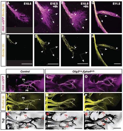
Visualizing ME and SA extension and selectively manipulating ME extension in mouse hindlimb. (A-H) Sequential extension of MEs (magenta) and SAs (yellow) beyond the sciatic plexus during development. Arrowheads: forefront SA growth cones extending along pre-extending MEs. Arrows (C-H): routing of MEs and SAs along dorsal and ventral nerve sheets. (I-Q) Dorsal wholemount view of MEs (I,L,O), SAs (J,M,P) or all axons (K, N,Q) in control (I-K) and Olig2Cre;Epha4fx/fx mouse embryos (L-Q) at E12.5. Dotted lines trace peroneal (PN) and solid lines tibial (TN) nerves. (J-P) Relative severity of ventral misrouting of MEs in Olig2Cre;Epha4fx/fx hindlimb (L,O) is faithfully mirrored by reduced/lost dorsal SA extension (M,P), also apparent by corresponding reduction/loss of pan-axon label (N,Q). Scale bars: 100 µm in B,D,F,H; 300 µm in K,N,Q. |