
Fig. 2
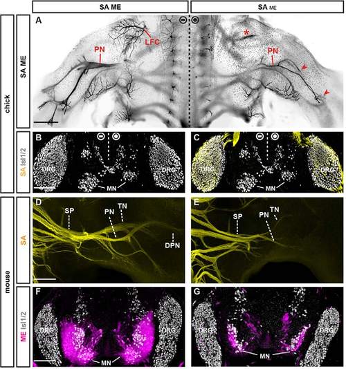
Effects of partial ME ablation on SA extension. (A) Dorsal whole-mount view of lumbar spinal cord and limbs in E6 chick embryo: peripheral axons visualized by anti-Tuj1 immunodetection (black). Severe reduction, but not loss, of crural (asterisk), peroneal (PN) and tibial nerves (TN) (arrowheads) upon unilateral transfection with a low titer (0.5µg/ml) of Hb9MN::Cre/PGKneolox2DTA plasmids. LFC, lateral femoral cutaneous nerve. (B,C) Transverse section of E6 chick spinal cord: partial ablation of motor neurons (MNs) after unilateral low-titer transfection. Anti-Isl1/2 immunofluorescence (gray) to label DRG neurons and MNs. Anti-TrkA immunofluorescence (yellow) to label DRG neurons. (D) Dorsal whole-mount view of SAs (yellow indicates Brn3atlz) at the sciatic plexus (SP) in E12.5 mouse embryo. DPN, deep peroneal nerve; PN, peroneal nerve; TN, tibial nerve. (E) Reduction, but not loss, of SAs beyond the sciatic plexus after delayed ablation of MEs in Olig2Cre;Isl2lxstopDTA embryo. (F) Transverse section of E12.5 control spinal cord: MNs labeled using Hb9MN::GFP (magenta). Anti-Isl1/2 immunofluorescence (gray) visualizes nuclei of DRG neurons and MNs. (G) Transverse section of E12.5 Olig2Cre;Isl2lxstopDTA spinal cord: severe reduction, but not complete absence, of MNs. Scale bars: 300μm in A; 100μm in B; 200μm in D,F. |