
Fig. 6
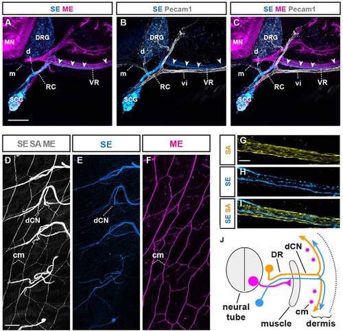
SEs extend along MEs and SAs to innervate dermis. (A-C) Transverse section of E14.5 mouse embryo at trunk levels: the majority of trunk-innervating SEs (arrowheads) extend along pre-extending axons (magenta indicates Hb9::eGFP) (A,C), rather than intersegmental blood vessels (vi) (gray indicates anti-Pecam1 immunofluorescence) (B,C). Subsets of SEs directly extend medially (m) and dorsally (d) along vasculature (B,C). (D-F) Dorsal whole-mount view of E18.5 mouse: dorsal cutaneous nerve (dCN) axons fanning out in trunk dermis and longitudinal projections by MEs into subdermal cutaneous maximus (cm) muscle (D,F). (E) SE projections (blue indicates anti-TH immunofluorescence) through dCN into dermis. (F) ME (magenta indicates Hb9::eGFP) innervation of subdermal cm muscle. (G-I) Longitudinal section through dCN at E18.5: separately labeled SEs (blue) and SAs (yellow indicates anti-TrkA immunofluorescence) can be seen. (J) Three axon types in dorsal ramus (ventral ramus, visceral or vascular trajectories are not depicted for simplicity). Magenta dots indicate cross-sections of cm MEs. DR, dorsal ramus; vi, intersegmental blood vessel; RC, ramus communicans; VR, ventral ramus. Scale bars: 150µm in A; 300µm in D; 50µm in G. |