
Fig. 5
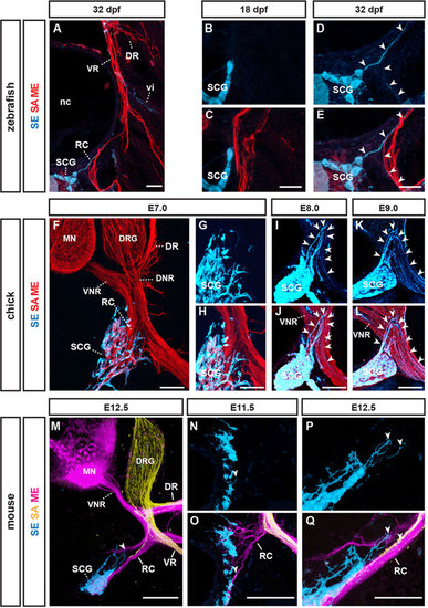
Conserved late extension of sympathetic efferent axons (SEs). (A) Transverse section of 32dpf zebrafish: SEs [blue, anti-tyrosine hydroxylase (TH) immunofluorescence] extending from sympathetic chain ganglion (SCG) along preformed peripheral nerves (red, SA/ME: anti-Tuj1 immunofluorescence). (B,C) TH+ SCG neurons prior to initiation SE axon extension at 18dpf. (D,E) Higher magnifications of A: TH+ SEs (arrowheads) extending along preformed peripheral axons. (F) Transverse sections of E7 chick embryo at trunk levels: TH+ SCG neurons (blue) around initiation SE extension relative to preformed PNs (red). (G,H) Magnified view of SCG neurons relative to preformed peripheral nerves. (I,J) TH+ SE axons begin extending from SCG along PNs at E8 (arrowheads). (K,L) SE axon advancing further peripherally along peripheral nerves at E9 (arrowheads). (M) Transverse section of E12.5 mouse embryo: TH+ SEs (arrowhead) beginning to extend from SCGs along pre-extending MEs and SAs in the ramus communicans (RC). (N,O) SCG and rc just prior to initiation of SE extension (arrowhead). (P,Q) Detailed view of SEs (arrowheads) extending along SAs and MEs of RC at E12.5. nc, notochord; DR/VR, dorsal/ventral ramus; vi, intersegmental blood vessel; DNR/VNR, dorsal/ventral nerve roots; RC, ramus communicans. Scale bars: 20µm in A,C,E; 100µm in F,H,J,L,M; 50µm in O,Q. |