
Fig. 5
- ID
- ZDB-FIG-140430-5
- Publication
- Gao et al., 2014 - Expression and functional characterization of Smyd1a in myofibril organization of skeletal muscles
- Other Figures
- All Figure Page
- Back to All Figure Page
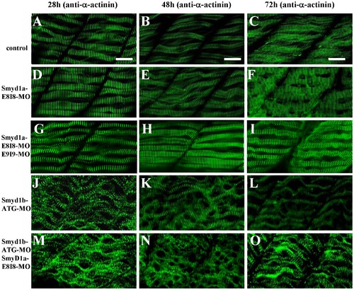
The effect of smyd1a or smyd1b single or double knockdown on the Z-line organization in skeletal muscles. Zebrafish embryos injected with smyd1b MO or smyd1a MO or both were fixed at 28, 48 and 72 hpf. Z-line organization was analyzed by immunostaining with anti-α-actinin antibody (EA-53), and followed by FTIC-labeled secondary antibody. The images represent side view of trunk muscles around segment 10. A–C. Lateral view of Z-line organization in skeletal muscle fibers of control-MO injected embryos at 28 (A), 48 (B) and 72 (C) hpf. D–F. Lateral view of Z-line organization in skeletal muscle fibers of smyd1a E8I8-MO injected embryos at 28 (D), 48 (E) and 72 (F) hpf. G–I. Lateral view of Z-line organization in skeletal muscle fibers of smyd1a E8I8-MO and E9I9-MO co-injected embryos at 28 (G), 48 (H) and 72 (I) hpf. J–L. Lateral view of Z-line organization in skeletal muscle fibers of smyd1b ATG-MO injected embryos at 28 (J), 48 (K) and 72 (L) hpf. M–O. Lateral view of Z-line organization in skeletal muscle fibers of smyd1a E8I8-MO and smyd1b ATG-MO co-injected embryos at 28 (M), 48 (N) and 72 (O) hpf. Scale bars: 20 μm in A–C. |
| Antibody: | |
|---|---|
| Fish: | |
| Knockdown Reagents: | |
| Anatomical Term: | |
| Stage Range: | Prim-5 to Protruding-mouth |
| Fish: | |
|---|---|
| Knockdown Reagents: | |
| Observed In: | |
| Stage Range: | Prim-5 to Protruding-mouth |