
Fig. 7
- ID
- ZDB-FIG-140430-7
- Publication
- Gao et al., 2014 - Expression and functional characterization of Smyd1a in myofibril organization of skeletal muscles
- Other Figures
- All Figure Page
- Back to All Figure Page
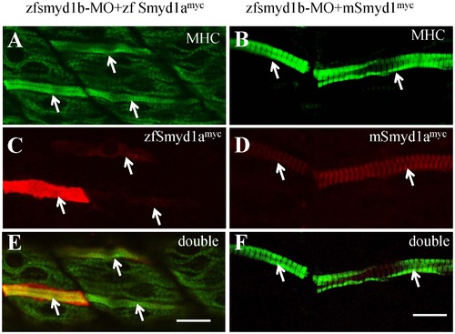
The myofibril defects from smyd1b knockdown could be rescued by ectopic expression of zebrafish smyd1a or mouse Smyd1. The smyd1b ATG-MO was co-injected with smyd1b:zfsmyd1amyc or smyd1b:mSmyd1myc transgene into zebrafish embryos at 1 or 2 cells stages. Myosin thick filament organization and transgene expression were analyzed by double immunostaining with anti-MHC (F59, green) and anti-myc (9E10, red) antibodies. A, C, E. Double immunostaining with anti-MHC (A) and anti-myc (C) antibodies shows the normal thick filament organization in myofibers expressing the myc-tagged zebrafish smyd1a transgene at 24 hpf. E, merged picture of A and C. B, D, F. Double immunostaining with anti-MHC (B) and anti-myc (D) antibodies shows the normal thick filament organization in myofibers expressing the myc-tagged mouse Smyd1 transgene at 24 hpf. F, merged picture of B with D. D and F showed the sarcomeric localization of myc-tagged mouse Smyd1 (red). The myc-tagged mouse Smyd1 was localized in the middle of the MHC thick filament (F), a region normally occupied by the M-line. Scale bars: 20 μm in E; 14 μm in F. |