
Fig. 4
- ID
- ZDB-FIG-140430-4
- Publication
- Gao et al., 2014 - Expression and functional characterization of Smyd1a in myofibril organization of skeletal muscles
- Other Figures
- All Figure Page
- Back to All Figure Page
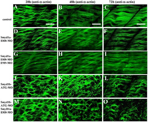
The effect of smyd1a or smyd1b single or double knockdown on thin filament organization in skeletal muscles. Zebrafish embryos injected with smyd1b MO or smyd1a MO or both were fixed at 28, 48 and 72 hpf. α-actin thin filament organization was analyzed by immunostaining with anti-α-actin antibody (Acl-20.4.2), and followed by FTIC-labeled secondary antibody. The images represent side view of trunk muscles around segment 10. A–C. Lateral view of thin filament organization in skeletal muscle fibers of control-MO injected embryos at 28 (A), 48 (B) and 72 (C) hpf. D–F. Lateral view of thin filament organization in skeletal muscle fibers of smyd1a E8I8-MO injected embryos at 28 (D), 48 (E) and 72 (F) hpf. G–I. Lateral view of thin filament organization in slow muscle fibers of smyd1a E8I8-MO and E9I9-MO co-injected embryos at 28 (G), 48 (H) and 72 (I) hpf. J–L. Lateral view of thin filament organization in skeletal muscle fibers of smyd1b ATG-MO injected embryos at 28 (J), 48 (K) and 72 (L) hpf. M–O. Lateral view of thin filament organization in skeletal muscle fibers of smyd1a E8I8-MO and smyd1b ATG-MO co-injected embryos at 28 (M), 48 (N) and 72 (O) hpf. Scale bars: 20 μm in A–C. |
| Antibody: | |
|---|---|
| Fish: | |
| Knockdown Reagents: | |
| Anatomical Term: | |
| Stage Range: | Prim-5 to Protruding-mouth |
| Fish: | |
|---|---|
| Knockdown Reagents: | |
| Observed In: | |
| Stage Range: | Prim-5 to Protruding-mouth |