
Fig. 1
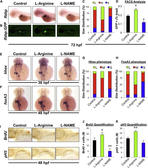
NO Signaling Regulates Liver Size during Development in Zebrafish (A) Effect of chemical modulators of NO signaling on liver formation. Zebrafish were exposed to chemicals (10 μM) from 24 to 72 hpf and subjected to in situ hybridization for the hepatocyte gene lfabp. Representative photomicrographs were taken at 10× magnification. (B) Effect of drug treatment on liver size in Tg(lfabp:GFP) embryos. Representative fluorescent photomicrographs were taken at 10× magnification. (C) Phenotypic analysis of liver size as determined by lfabp in situ hybridization in treated embryos at 72 hpf (S, small; M, medium; L, large, n > 50 embryos per treatment). (D) Effect of drug treatment on the percentage of hepatocytes specified during liver formation. Chemically treated Tg(lfabp:GFP) embryos were dissociated and the percentage of GFP positive hepatocytes was analyzed by fluorescence-activated cell sorting (FACS). n = 4; ANOVA, p < 0.05 in comparison to control. (E and F) Effect of drug treatment on hepatic progenitor cells. Zebrafish embryos were exposed to chemicals (10 μM) from 18 to 36 hpf or 24–48 hpf and subjected to in situ hybridization for the endodermal genes hhex and foxA3, respectively. (G and H) Phenotypic analysis of hepatic bud size as determined by hhex and foxA3 in situ hybridization in treated embryos at 36 and 48 hpf, respectively (n > 25 embryos per treatment). (I) Effect of drug treatment on hepatic progenitor cell proliferation as determined by BrdU incorporation or pH 3 staining, respectively. (J and K) Quantification of hepatic progenitor cell proliferation. Representative photomicrographs were taken and cell counts were performed. n = 10; ANOVA, p = 0.042, p < 0.001 in comparison to control. Results in (D), (J), and (K) show mean ± SEM. |
| Genes: | |
|---|---|
| Fish: | |
| Condition: | |
| Anatomical Term: | |
| Stage Range: | Prim-25 to Protruding-mouth |
| Fish: | |
|---|---|
| Condition: | |
| Observed In: | |
| Stage Range: | Prim-25 to Protruding-mouth |