
Fig. 3
- ID
- ZDB-FIG-140115-19
- Publication
- Kim et al., 2013 - A Complex of BBS1 and NPHP7 Is Required for Cilia Motility in Zebrafish
- Other Figures
- All Figure Page
- Back to All Figure Page
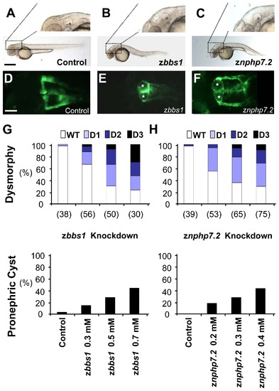
Depletion of zBbs1 and zNphp7.2 causes dose-dependent hydrocephalus and pronephric cysts. Zebrafish embryos injected with MOs at the 1-cell stage were evaluated for their phenotype at 55 hpf. (A) Control zebrafish embryo with magnified normal brain area. Scale bar = 500 μm. (B and C) zbbs1 AUG MO-injected embryo and znphp7.2 SP1 MO-injected embryo with hydrocephalus. (D) The Tg(WT1b:EGFP) transgenic line shows normal pronephric glomerulus and tubules (Scale bar = 100 μm), (E and F) zbbs1 AUG MO-injected embryo and znphp7.2 SP1 MO-injected embryo show pronephric cysts (asterisks). At 55 hpf, larval dysmorphy was categorized according to the visual scale (Fig. S3). The degree of dysmorphy was increased with the amount of injected MOs (G, zbbs1 AUG MO; H, znphp7.2 SP1 MO). (G and H) Cystic pronephric phenotypes were dose-dependent in zebrafish embryos injected with zbbs1 and znphp7.2 MO respectively. The graphs show percentages of the number (n) of embryos that were examined. |
| Gene: | |
|---|---|
| Fish: | |
| Knockdown Reagents: | |
| Anatomical Terms: | |
| Stage: | Long-pec |
| Fish: | |
|---|---|
| Knockdown Reagents: | |
| Observed In: | |
| Stage: | Long-pec |