
Fig. 7
- ID
- ZDB-FIG-140115-24
- Publication
- Kim et al., 2013 - A Complex of BBS1 and NPHP7 Is Required for Cilia Motility in Zebrafish
- Other Figures
- All Figure Page
- Back to All Figure Page
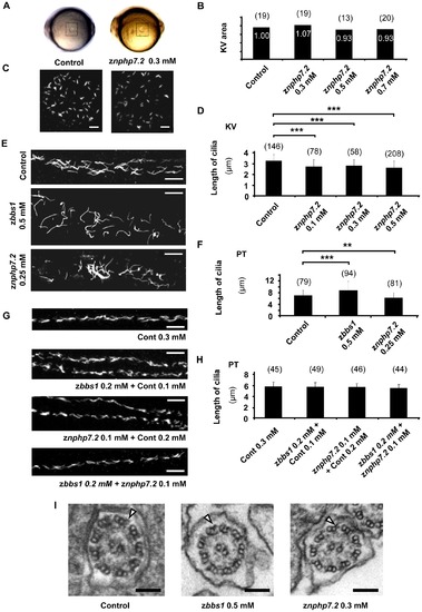
Knockdown of znphp7.2 showed normal development of cilia. (A) Images of live zebrafish embryos at the 8–10 somite stage. Kupffer′s vesicle is located in the dashed box. (B) Measurement of relative KV area with setting control as ‘1.00’. The knockdown of znphp7.2 did not significantly affect on KV development and area. (C) Images of cilia in the KV of 8–10 somite stage were stained for acetylated tubulin. Scale bar = 10 μm. (D) zNphp7.2-deficient embryos showed shortened length of cilia in KV compared to control embryos. (E) Staining of acetylated tubulin in the anterior pronephric tubule of morphants at 48 hpf displayed that the overall distribution of cilia remained unchanged compared to control (Scale bar = 10 μm) even though (F) the knockdown either of zbbs1 showed longer and the knockdown of znphp7.2 showed shorter cilia. (PT, Pronephric Tubule) (G and H) The morphology and length of cilia in posterior pronephric tubule appeared unchanged in combined knockdown of zbbs1 and znphp7.2 compared to single knockdown of zbbs1 or znphp7.2 or compared to Cont MO injected embryos. (PT, Pronephric Tubule) (I) The morphants of zbbs1 and znphp7.2 displayed normal ultrastructure of motile cilia in pronephric tubule without recognizable deficiency in dynein arms (outer dynein arm marked by arrowhead). The numbers (n) in the graphs are the number of total cilia which were examined. 4–6 individual embryos per group were examined. |
| Fish: | |
|---|---|
| Knockdown Reagents: | |
| Observed In: | |
| Stage Range: | 5-9 somites to Long-pec |