
Fig. S8
- ID
- ZDB-FIG-140115-30
- Publication
- Kim et al., 2013 - A Complex of BBS1 and NPHP7 Is Required for Cilia Motility in Zebrafish
- Other Figures
- All Figure Page
- Back to All Figure Page
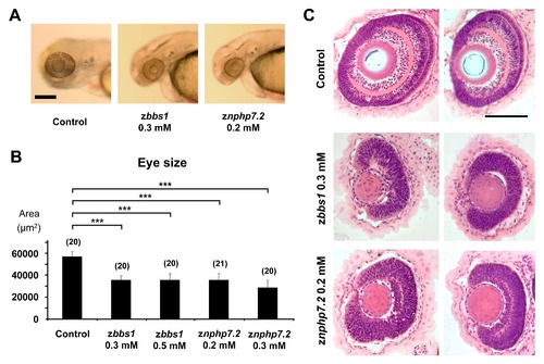
zbbs1 and znphp7.2 morphants display reduced eye size and defective retinal layer formation. The eye size as area (μm2) was measured for control embryos, zbbs1 and znphp7.2 morphant embryos at 80 hpf. (A) Representative brightfield images showing reduced eye size for zbbs1 and znphp7.2 morphants in comparison to the control (Scale bar = 200 μm). (B) Statistical quantification of the measurements proved that the reduction in eye size for zbbs1 and znphp7.2 morphants was significant in comparison to the control. (C) Histological cross-sections of 96 hpf zbbs1 and znphp7.2 morphants revealed defective layer formation in comparison to the control. Two representative images are shown for each setting. (Scale bar = 100 μm). |
| Fish: | |
|---|---|
| Knockdown Reagents: | |
| Observed In: | |
| Stage Range: | Protruding-mouth to Day 4 |