
Fig. 5
- ID
- ZDB-FIG-140114-59
- Publication
- Leigh et al., 2013 - Mmp17b is essential for proper neural crest cell migration in vivo
- Other Figures
- All Figure Page
- Back to All Figure Page
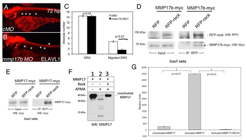
RECK blocks MMP17 activity in vitro. Elavl1 staining of control MO (cMO) (A) or mmp17b MO1 (B) injected 72 hpf embryos. Asterisks show the location of DRGs (dorsal root ganglia). Mmp17b knockdown embryos lack proper DRG development compared to controls. Quantitation of number of DRGs and migrated DRGs are indicated in panel C. Panel D is immunoprecipitation of myc-Mmp17b and RFP-Reck in Cos7 cells. Left panels indicate input of the two proteins in the sample, and right panels are RFP antibody immunoprecipitated samples followed by western blot. Arrow shows the pull down Mmp17b protein. Panel E is immunoprecipitation of myc-MMP17 and RFP-Reck in Cos7 cells. Left panel indicates input of MMP17 proteins in the lysate and the right panels indicate RFP antibody immunoprecipitated samples followed by western for myc epitope. Panel F is western blot of myc-MMP17 unactivated and activated with 4-aminophenylmercuric acetate (APMA) using MMP17 antibody. Activated MMP lanes show multiple bands, which are absent in samples co-incubated with RECK protein. Panel G is FRET-based fluorescence readout for MMP activity using the samples run on western blots in panel F. |
| Antibody: | |
|---|---|
| Fish: | |
| Knockdown Reagent: | |
| Anatomical Term: | |
| Stage: | Protruding-mouth |
| Fish: | |
|---|---|
| Knockdown Reagent: | |
| Observed In: | |
| Stage: | Protruding-mouth |