
Fig. 2
- ID
- ZDB-FIG-140114-56
- Publication
- Leigh et al., 2013 - Mmp17b is essential for proper neural crest cell migration in vivo
- Other Figures
- All Figure Page
- Back to All Figure Page
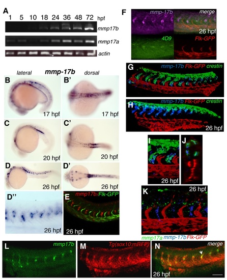
Spatial and temporal characterization of mmp17b expression. A is RT-PCR showing temporal expression of mmp17b and mmp17a compared to β-actin. Mmp17b expression commences at around 18 hpf while mmp17a is expressed as early as 5 hpf. B-E are whole mount in situ hybridization (WISH) panels of mmp17b at different embryonic stages (B and B′, 17 hpf; C and C′, 20 hpf; D and D′, 26 hpf). Earlier in development mmp17b is expressed more dorsally and then moves more posterior and ventral as development continues. Panel E shows mmp17b fluorescent in situ hybridization (FISH) in red and immunofluorescence (IF) for Flk-GFP in a 26 hpf embryo, which illustrates lack of mmp17b expression in the vasculature. Panels F, G-H, I-J and K are three color WISH staining performed as described in materials and methods with different probes as indicated in each panel. In panel F, mmp17b (purple), lower left is 4D9 staining (green) the muscle pioneer cells, lower right is Flk-GFP marking (red) the endothelial cells, and upper right is a merge. In this panel you can observe that 4D9 staining is located in the same region as mmp17b. G-H panel shows three color ISH image of 26 hpf zebrafish trunk and plexus with mmp17b (blue), Flk-GFP (red), and crestin (green). In the trunk image, crestin and mmp17b are overlapping in expression suggesting co-expression of these two genes in the same cell. The expression of both crestin and mmp17b are more dorsal in posterior regions of the embryo. Panels I-J represent high-powered image of the panel G. J is an optical section of panel G. Panel K is a three color image of 26 hpf zebrafish trunk with mmp17a (green), mmp17b (blue), and Flk-GFP (red). This image illustrates that mmp17a and mmp17b are expressed in different regions of the developing embryo. B-D are lateral views, B’-D’ are dorsal views. D” is a close-up of the panel D. Panels L-N are single plane confocal images of a 26 hpf embryo stained for mmp17b using FISH (panel L, green), and immunostaining for sox10 cells labelled with RFP (panel M, red). Panel N is the merged images showing co-localization of mmp17b and sox10 as indicated by yellow color (arrowhead). |
| Genes: | |
|---|---|
| Antibody: | |
| Fish: | |
| Anatomical Terms: | |
| Stage Range: | 4-cell to Protruding-mouth |