
Fig. 3
- ID
- ZDB-FIG-140114-57
- Publication
- Leigh et al., 2013 - Mmp17b is essential for proper neural crest cell migration in vivo
- Other Figures
- All Figure Page
- Back to All Figure Page
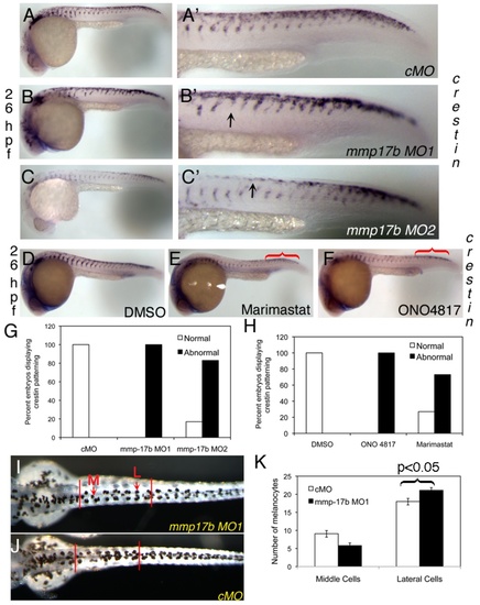
Mmp17b is involved in neural crest patterning. A-C and D-F are WISH staining for crestin in control MO (cMO) (A), MO1 (B), and MO2 (C) injected 26 hpf or DMSO (D), Marimastat (E) and ONO-4817 (F) treated 26 hpf embryos. A′-C′ are high powered images of the trunk regions of A-C. Arrowheads indicate crestin+ cells misplaced in the trunk. There is a mis-patterning of crestin in the trunk of the MO1 and MO2 injected embryos compared to control. There is also an accumulation of crestin+ cells in the posterior of the embryo (white bracket) compared to control. This is quantitated in panel G. N=25 for cMO; n=19 for MO1; n=18 for MO2. In panels D-F, WISH staining for crestin in MMP inhibitor treated 26 hpf embryos shows mis-patterning similar to mmp17b KD embryos (A-C). An accumulation of crestin+ cells in the posterior of the MMP inhibitor treated embryos is also observed (red brackets). This is quantitated in panel H. N=12 for DMSO; n=8 for ONO 4817; n=10 for Marimastat. I-K shows melanocyte quantitation done on 72 hpf fish. Dorsal images were taken of 72 hpf fish injected with either control MO (J) or mmp17b MO1 (I). The number of medial (M, red arrow) and lateral (L, red arrow) melanocytes is counted between the two vertical bars illustrated in panels I and J for 10 fish in each category. The results were quantitated in panel K. The medial cells were not statistically different but the lateral cells were at a p-value of less than 0.05. |
| Gene: | |
|---|---|
| Fish: | |
| Knockdown Reagents: | |
| Anatomical Term: | |
| Stage: | Prim-5 |
| Fish: | |
|---|---|
| Knockdown Reagents: | |
| Observed In: | |
| Stage Range: | Prim-5 to Protruding-mouth |