
Fig. S8
- ID
- ZDB-FIG-130816-8
- Publication
- Minchin et al., 2013 - Oesophageal and sternohyal muscle fibres are novel Pax3-dependent migratory somite derivatives essential for ingestion
- Other Figures
- All Figure Page
- Back to All Figure Page
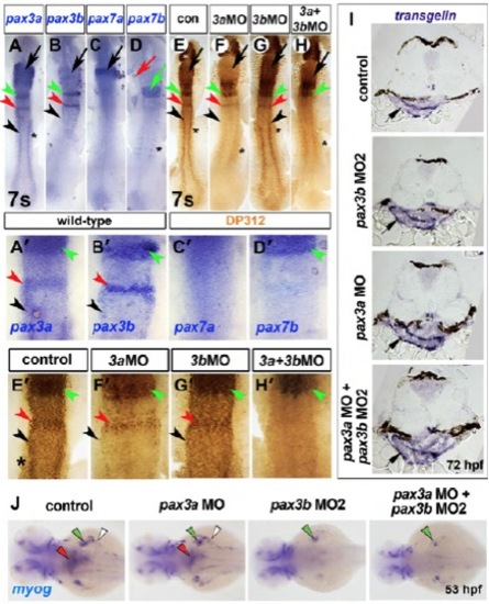
pax3b morpholinos specifically reduce Pax3b protein but not transgelin expression. In situ mRNA hybridization for pax3a (A,A′), pax3b (B,B′), pax7a (C,C′), pax7b (D,D′), transgelin (I), myog (J) and parallel immunodetection of nuclear Pax3/7 protein (brown, DP312 antibody, E-H′) in 7 ss embryos injected with the indicated MOs. Dorsal view flatmounts with anterior towards the top (A-H′) or left (J). Transverse cryosections with dorsal towards the top (I). (A-B′) pax3a was detected in rhombomeres 2 (green arrowheads, A,A′) and 4 (red arrowheads, A,A′), lateral neural plate (black arrowheads, A,A′) and somites (asterisk, A,A′). Concurrently, pax3b mRNA was also observed in rhombomere 2 (green arrowhead, B,B′), rhombomere 4 (red arrowhead, B,B′) and lateral neural plate (black arrowhead, B,B′) but was lacking in somites (B,B′). (C-D′) The related pax3/7 family member pax7a was expressed solely in the midbrain region (black arrow, C), whereas pax7b mRNA was detected in somites (asterisk, D) and a broad anterior hindbrain region (green arrow and arrowhead, D,D2). Therefore, of pax3/7 family members, pax3a was the most widely and abundantly expressed at 7 ss, encompassing expression domains of all other pax3/7 family members. (E,E′) Pax3/7 immunoreactivity closely resembles the sum of all pax3/7 mRNA localisation, with the addition of signal in the forming retina owing to the known reaction of DP312 with Rx proteins (asterisks, E) (Davis et al. 2005). (F-G′) Injection of MO1 specific to pax3b did not result in a detectable reduction of Pax3/7 protein (G,G′), in agreement with residual reactivity to other Pax3/7 proteins, particularly Pax3a. After MO1-mediated Pax3a reduction (F,F′), Pax3/7 protein is clearly evident in regions of pax3b expression including rhombomere 2 (F,F′, green arrowheads), rhombomere 4 (F,F′, red arrowheads) and lateral neural plate (F,F′, black arrowheads), suggesting that residual Pax3/7 reactivity represents Pax3b. Using two non-overlapping pax3a MOs, we failed to observe the head phenotype previously described for pax3a knockdown (Lin et al. 2009). (H,H′) Knockdown of both Pax3b and Pax3a proteins resulted in the ablation of the residual Pax3/7 immunoreactivity in rhombomere 4 and lateral neural plate, which corresponded to Pax3b. Remaining Pax3/7 protein anteriorly is likely to reflect Pax7b protein (green arrowhead, H′) (Davis et al. 2005; Minchin and Hughes 2008). Therefore, we conclude that residual immunoreactivity after Pax3a reduction corresponds in part to Pax3b protein and that this is specifically reduced by pax3b MO1, which does not itself target Pax3a. Similar results were obtained with pax3a MO2 and pax3b MO2 (Table S1). (I) transgelin mRNA in oesophagus at 72 hpf did not change after pax3 manipulation (arrows). (J) myog mRNA in oesophagus (red arrowheads), pectoral fin muscle (green arrowheads) and posterior hypaxial muscles (white arrowheads) is reduced in pax3b and pax3a+pax3b double morphants. |