
Fig. 1
- ID
- ZDB-FIG-130816-12
- Publication
- Minchin et al., 2013 - Oesophageal and sternohyal muscle fibres are novel Pax3-dependent migratory somite derivatives essential for ingestion
- Other Figures
- All Figure Page
- Back to All Figure Page
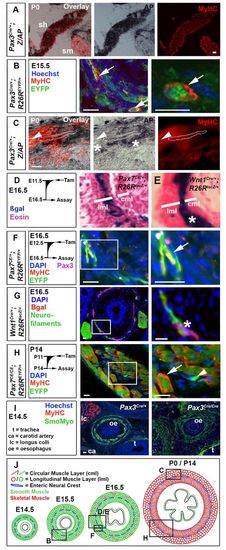
Pax3- and Pax7-expressing cells contribute to mouse OSM. (A-I) Immunodetection of MyHC (A-C,F,H,I), smooth muscle myosin (I), EYFP (B,F,H) and βgal (G); nuclei staining (DAPI/Hoechst; B,F-I); and histochemistry for alkaline phosphatase (AP; A,C) or βgal (D,E) on transverse cryosections of murine oesophagus. (A) The sternohyoid muscle (sh) was AP+, whereas the adjacent sternomastoid muscle (sm) was unlabelled. (B) Pax3Cre/+;R26REYFP mice show MyHC in some EYFP+ cells (arrows) at E15.5. (C) By P0, Pax3Cre/+;Z/AP mice have OSM cells marked with AP (dots outline an area of staining over a fibre). Some OSM is unmarked (arrowheads), and neural cells between the longitudinal and circumferential OSM layers are marked (asterisks). (D) Tamoxifen treatment of Pax7CE/+;R26RlacZ mice at E11.5 marked cells at E16.5 in outer longitudinal muscle layer (lml, where OSM is present) but only rare cells in inner circumferential muscle layer [cml, lacking OSM at this stage (see A)]. (E) Wnt1Cre;R26RlacZ (right panel) mice had neural crest cells between the muscle layers (asterisk). (F) Tamoxifen treatment of Pax7CE/+;R26REYFP mice at E12.5 marked OSM cells in lml at E16.5 with EYFP (arrow). Small rare Pax3+ cells are found adjacent to the OSM. (G) At E16.5, Wnt1Cre;R26RlacZ mice have βgal in a thin layer of neurofilament-containing cells between the muscle layers (asterisk). (H) Treatment of Pax7CE/CE;R26REYFP mice with tamoxifen at P11 led to abundant EYFP-marked OSM cells at P14 (arrow) and smaller EYFP-marked cells lacking MyHC (arrowhead). (I) At E14.5, Pax3Cre/Cre mice show slightly reduced and disorganised smooth muscle myosin (green), compared with Pax3Cre/+ littermates. There is a lack of longus colli striated muscle (lc) in mutant. ca, carotid artery; t, trachea; oe, oesophagus. (J) Mouse OSM development (red, sarcomeric myosin) among smooth muscle (green, smooth myosin), drawn approximately to scale. Boxed areas in the left panels are magnified on the right. Boxed areas in J are shown in panels above. Scale bars: 50 μm in A; 20 μm in B-I. |