
Fig. S11
- ID
- ZDB-FIG-130816-5
- Publication
- Minchin et al., 2013 - Oesophageal and sternohyal muscle fibres are novel Pax3-dependent migratory somite derivatives essential for ingestion
- Other Figures
- All Figure Page
- Back to All Figure Page
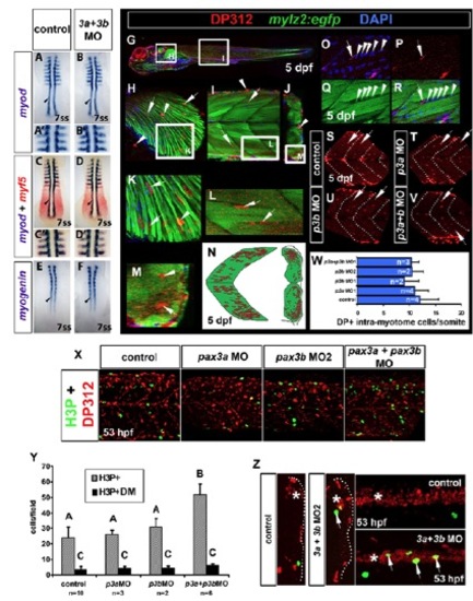
Early myogenesis and Pax3/7+ intra-myotome cells occur normally in pax3b morphants. In situ hybridisation of myod (A-B&prime& blue, C-D′), myf5 (red, C-D′) and myogenin (E,F). Immunodetection of DP312 (G-M,O-V; red, X,Z) and phospho-histone H3 (H3P) (green, X,Z) in wild-type (A-F,X,Z) and Tg(mylz2:egfp) backgrounds. (A-F) Expression of myod, myf5 and myog at 7 ss are unaffected in pax3a+pax3b double morphants. (G-M) DP312+ intra-myotome cells (IMCs) localise to inter-fibre regions, reminiscent of Pax7+ myogenic cells (Seger et al., 2011). (N) IMC location. (O-R) IMCs are likely to be myogenic as they are observed closely juxtaposed to myogenic nuclei (Q,R, arrowheads) during myotube fusion (R). (S-W) Number of IMCs at 5 dpf is unchanged in pax3a, pax3b and pax3a+pax3b double morphants. (X,Y) Cell proliferation of dermomyotome cells (DM), as assessed by faint DP312/H3P+ nuclei (green, X) is unchanged after pax3 manipulation (Y). However, total H3P+ nuclei are significantly increased in pax3a+pax3b double morphants (Y) (P<0.0001). (Z) Increase in H3P+ nuclei occurs in the neural tube. Asterisks indicate neural tube. Arrows indicate H3P+ nuclei within neural tube. One-way ANOVA and Tukey’s post hoc test. |