
Fig. S4
- ID
- ZDB-FIG-130816-10
- Publication
- Minchin et al., 2013 - Oesophageal and sternohyal muscle fibres are novel Pax3-dependent migratory somite derivatives essential for ingestion
- Other Figures
- All Figure Page
- Back to All Figure Page
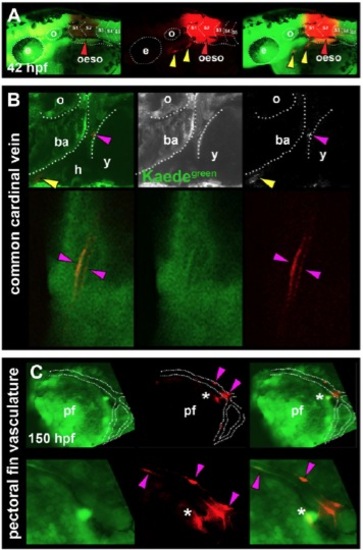
Cells associated with the heart and pectoral fin vasculature are derived from anterior somites in zebrafish. Primary green Kaede (Kaedegreen) fluorescence and photoconverted red Kaede (Kaedered) fluorescence. All images are lateral view wholemounts with anterior towards the left and dorsal towards the top. (A) Kaedered fluorescence localised to the oesophagus after photoconversion at 18 ss (red arrowheads). SHM was also labelled with Kaedered (yellow arrowheads). (B) Photoconversion of S2 at 14-18 ss marks vascular elements associated with the common cardinal vein (magenta arrowheads) and SHM (yellow arrowheads). (C) In addition, cells associated with pectoral fin vasculature and other unidentified cells were labelled after photoconversion of S1/2 (magenta arrowheads and asterisks, respectively). Labelled blood vessels were observed only in tissues adjacent to the original somites converted, suggesting regional restriction of somite-derived vasculature. ba, branchial arches; oeso, oesophagus; h, heart; pf, pectoral fin; o, otic vesicle; y, yolk. |