|
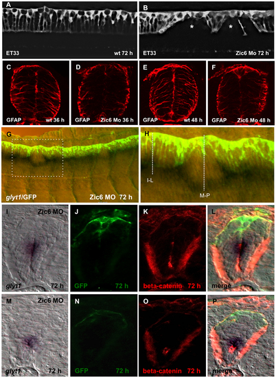
Zic6 is required for development of the radial glial scaffold and attachment of RP cells to the dorsal surface of central canal. Confocal images, lateral view of the spinal cord, 72 hpf. SqET33, control (A) and Zic6 morphant (B). The asterisk indicates the “gaps” in the palisade of RP cells. (C–F), Immunostaining of the transverse sections of the spinal cord with anti-GFAP antibody. (G), Double whole-mount in situ hybridization (glyt1, dark purple) and immunostaining (GFP, yellow/green) of the spinal cord Zic6 morphant. The dashed rectangular marks the magnified region (H). The dashed lines show the approximate positions for the transverse sections shown in (I–P). (I–P), distribution of glyt1, β-catenin and GFP in two positions in the spinal cord of Zic6 morphants shown in H, corresponding to I–L and M–P.
|