
Fig. 5
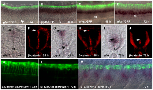
Growth of the RP processes correlates with a shift of glyt1 expression along D–V axis. (A-D), Double whole mount in situ hybridization (glyt1, dark purple) and immunostaining (GFP, green). glyt1 is expressed in the midline glial cells except RP; GFP is expressed in the RP cells. (E-J), Whole mount in situ hybridization (glyt1, dark purple) and immunostaining (β-catenin, red), the transverse sections of the spinal cord. The arrow shows an approximate position of attachment of the RP process to the apical surface of central canal. Confocal images of the spinal cord of pard6γb heterozygote (K) and pard6γb mutant (L) transgenic fish. (M), double whole mount in situ hybridization (glyt1, dark purple) and immunostaining (GFP, green) of pard6γb mutant. Abbreviation: fp, floor plate. |
| Genes: | |
|---|---|
| Antibody: | |
| Fish: | |
| Anatomical Terms: | |
| Stage Range: | Prim-5 to Protruding-mouth |
| Fish: | |
|---|---|
| Observed In: | |
| Stage: | Protruding-mouth |