
Fig. 1
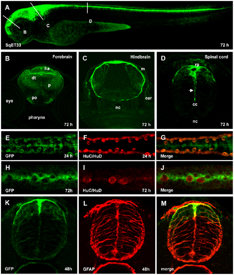
Characterization of SqET33 transgenic line. (A), Confocal image of 3 dpf larva of SqET33 line, lateral view. Dashed lines depict the position of transverse sections shown in B–D. (B–D), transverse sections, immunofluorescent staining with anti-GFP antibodies. Arrow indicates the elongated central process of the roof plate cell. (E–J), whole-mount immunofluorescent staining with anti-GFP (midline RP cells and lateral dorsal interneurons) and anti-HuC/HuD (lateral dorsal neurons) antibodies, dorsal view of spinal cord. (K–M), immunofluorescent staining with anti-GFP (green) and anti-GFAP (red) antibodies, transverse section of the spinal cord. Abbreviations: cc, central canal; dt, dorsal thalamus; ha, habenula; m, meninx; nc, notochord; p, pallium; po, preoptic area; rp, roof plate. |
| Gene: | |
|---|---|
| Antibodies: | |
| Fish: | |
| Anatomical Terms: | |
| Stage Range: | Prim-5 to Protruding-mouth |