
FIGURE
Fig. 3
Fig. 3
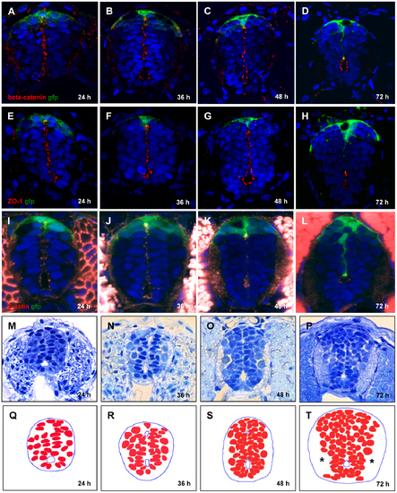
Conversion of primitive lumen into central canal. (A–L), Contraction of opposed apical surfaces reflects recession of the primitive lumen into a central canal between 24 and 72 hpf. Immunofluorescent staining by a combination of anti-GFP with anti-β-catenin antibody (A–D), anti-ZO-1 antibody (E–H), and phalloidin that detects F-actin (I–L). Increase in cell number and build-up of axonal tracts (*), plastic transverse sections of the spinal cord (M–P). (Q–T), Schematics corresponding to (M–P) showing cell nuclei. |
Expression Data
| Gene: | |
|---|---|
| Antibody: | |
| Fish: | |
| Anatomical Terms: | |
| Stage Range: | Prim-5 to Protruding-mouth |
Expression Detail
Antibody Labeling
Phenotype Data
Phenotype Detail
Acknowledgments
This image is the copyrighted work of the attributed author or publisher, and
ZFIN has permission only to display this image to its users.
Additional permissions should be obtained from the applicable author or publisher of the image.
Full text @ PLoS One