|
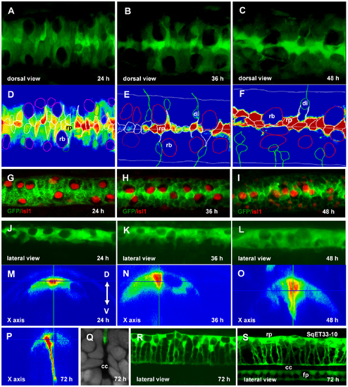
Re-orientation of the RP cells. Confocal images of the spinal cord of SqET33 line at different developmental stages (A–C, dorsal view, J–L, R, lateral view, M–P, orthogonal optical sections) and outline of the dorsal cells (D–F) superimposed onto the GFP intensity chart. Note that RB cells undergo the lateral-medial displacement. (G–I), Whole-mount immunohistochemistry detecting Islet1 in RB cells (red, nuclei), dorsal view of the spinal cord. (Q), Transverse section of the spinal cord at high magnification showing a fine structure of the central canal. RP process is stained with anti-GFP (green) and nuclei are counterstained with DAPI (grey). (S), Confocal image of the spinal cord of SqET33-10 line expressing GFP in the roof and floor plates, lateral view. Abbreviations: cc, central canal; di, dorsal interneurons; fp, floor plate; rb, Rohon-Beard cells; rp, roof plate cells.
|