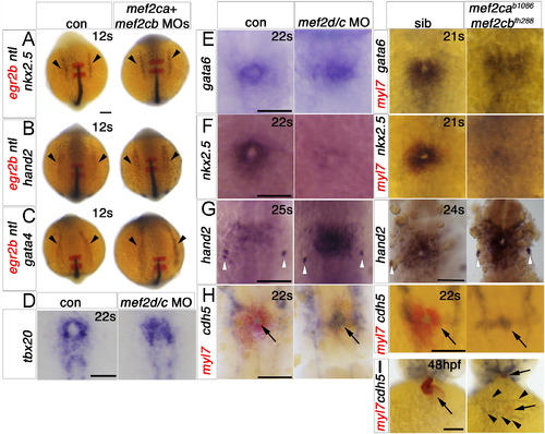
Cardiomyocytes of mef2ca;mef2cb dual loss of function are specified but developmentally arrested. In situ mRNA hybridisation for indicated genes (wholemounts in dorsal view, anterior to top, except J, ventral view). A–C. ALPM expression (arrowheads) of nkx2.5 (A), hand2 (B) and gata4 (C) at 12 ss is unaffected by lack of Mef2ca and Mef2cb. Notochord and rhombomeres 3 and 5 are marked by ntl (blue) and egr2b (red), respectively. D,E.Tbx20 and gata6 mRNAs are present but disorganised in the heart region of mef2d/c morphants and mef2cab1086;mef2cbfh288 mutant embryos (lacking myl7 expression in red, E, right panel) compared to the typical ring-shape in control and sibling embryos. F. During heart cone stage (22 ss), nkx2.5 mRNA is abolished. G. hand2 mRNA is enhanced in a sheet of cells spanning the cardiac region but not elsewhere. Pharyngeal pouch expression is unchanged (white arrowheads). H, Whereas myocardial cells are undifferentiated in mef2d/c morphants and mef2cab1086;mef2cbfh288 mutants (myl7, red), the endocardium is expanded and cdh5 is up-regulated. I. Endocardium (cdh5, arrow) lines the myocardium (myl7, red) in the normal looped heart of a sibling embryo. In mef2ca;mef2cb mutant embryos, little endocardial makrer is co-localised with the residual myocardium (arrows). Extra cdh5-expressing tissue is detected in the cardiac region (arrowheads). Scale=100 μm. (For interpretation of the references to color in this figure legend, the reader is referred to the web version of this article.)
|
