
Fig. 2
- ID
- ZDB-FIG-121030-32
- Publication
- Hinits et al., 2012 - Zebrafish Mef2ca and Mef2cb are essential for both first and second heart field cardiomyocyte differentiation
- Other Figures
- All Figure Page
- Back to All Figure Page
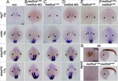
Early cardiomyocytes fail to differentiate after loss of Mef2c function. A–F. In situ mRNA hybridisation for myl7, vmhc and smyd1b in wild type control (A), mef2cab1086+mef2cb MO (B), mef2cab1086 (C) mef2cb MO (D), mef2cbfh288 (E) and mef2cab1086;mef2cbfh288 (F) embryos, shown in wholemounts in dorsal view, anterior to top. Loss of both mef2ca and mef2cb function greatly reduces myl7, vmhc and smyd1b mRNAs in the bilateral heart fields (arrowheads; A,B and F). Mef2cab1086 mutant embryos have weak myl7 and smyd1b mRNAs early, but recover later, and show no change in vmhc (C). Mef2cb single morphants or mef2cbfh288 mutant show no changes (D). G. Immunostaining for MyHC (A4.1025) in 24 ss mef2cab1086;mef2cbfh288 embryos and their siblings, shown in wholemounts in dorsal view, anterior to top (left panel) and lateral view, anterior to left (right panel). No MyHC is detected in the heart, whereas somitic muscle appears normal (white arrowheads). Scale=100 μm. |
| Genes: | |
|---|---|
| Antibody: | |
| Fish: | |
| Knockdown Reagent: | |
| Anatomical Terms: | |
| Stage Range: | 10-13 somites to 20-25 somites |
| Fish: | |
|---|---|
| Knockdown Reagent: | |
| Observed In: | |
| Stage Range: | 10-13 somites to 20-25 somites |
Reprinted from Developmental Biology, 369(2), Hinits, Y., Pan, L., Walker, C., Dowd, J., Moens, C.B., and Hughes, S.M., Zebrafish Mef2ca and Mef2cb are essential for both first and second heart field cardiomyocyte differentiation, 199-210, Copyright (2012) with permission from Elsevier. Full text @ Dev. Biol.