FIGURE

Fig. S2

ID
ZDB-FIG-121030-23
Publication
Hinits et al., 2012 - Zebrafish Mef2ca and Mef2cb are essential for both first and second heart field cardiomyocyte differentiation
Other Figures
All Figure Page
Back to All Figure Page
Fig. S2

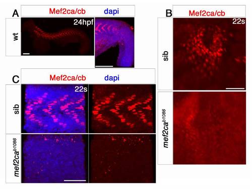
Mef2c protein is downregulated in mef2ca mutants.
Confocal stacks of Mef2ca/cb immunodetection (Anaspec, red in A-C) and DAPI (blue, A and C) in wildtype and mef2ca mutant embryos. A. Reaction with Mef2c antibody is strong in nuclei of differentiated slow muscle fibres in the somite (left panel), but is missing from the PSM (right panel), and from cranial ganglia indicating that the antibody is not crossreacting with Mef2d and Mef2a protein, respectively. B and C. Anti-Mef2c antibody reacts weakly in CMs of mef2cab1086 mutant embryos (B), but is absent from nuclei in the myotome of the somites (C). Scale = 50 μm (except in A=100 μm).

Expression Data
Antibody:
Fish:
Anatomical Terms:
Stage Range: 20-25 somites to Prim-5

Expression Detail
Antibody Labeling
Phenotype Data

Phenotype Detail
Acknowledgments
This image is the copyrighted work of the attributed author or publisher, and ZFIN has permission only to display this image to its users. Additional permissions should be obtained from the applicable author or publisher of the image.

Reprinted from Developmental Biology, 369(2), Hinits, Y., Pan, L., Walker, C., Dowd, J., Moens, C.B., and Hughes, S.M., Zebrafish Mef2ca and Mef2cb are essential for both first and second heart field cardiomyocyte differentiation, 199-210, Copyright (2012) with permission from Elsevier. Full text @ Dev. Biol.