
FIGURE
Fig. S2
Fig. S2
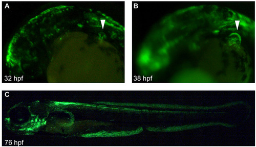
Distribution of gal4 reporter in the hspGFFDMC131A line. The hspGFFDMC131A line was crossed with the UAS:GFP line; Gal4 distribution is shown by GFP fluorescence. (A-C) Lateral view of embryos at 32 (A), 38 (B) and 76 (C) hpf. At 32 hpf, GFP was distributed in the epidermis, brain and AER (arrowhead in A). At 38 hpf, the AF was clearly visible by the GFP distribution (arrowhead in B). (C) At 76 hpf, GFP was distributed in several organs (brain, branchiae, spinal cord, AF and median fin fold). |
Expression Data
Expression Detail
Antibody Labeling
Phenotype Data
Phenotype Detail
Acknowledgments
This image is the copyrighted work of the attributed author or publisher, and
ZFIN has permission only to display this image to its users.
Additional permissions should be obtained from the applicable author or publisher of the image.
Full text @ Development