
FIGURE
Fig. S5
Fig. S5
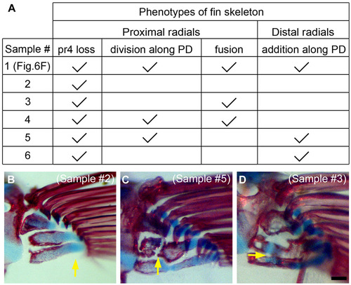
Phenotypes of fin skeleton after AF removal three times. (A) A table showing phenotype of radials in all six samples in which we observed a skeletal pattern. Sample 1 is the one that is shown in Fig. 6F. Phenotypes are categorized into proximal radials (loss, division, and fusion) and distal radials (addition). (B-D) Skeletal patterns in three other specimens are shown. At the points indicated by yellow arrows, pr (proximal radial) 4 is lost (B), pr2 is divided (C), or pr2 and pr3 are fused (D). |
Expression Data
Expression Detail
Antibody Labeling
Phenotype Data
Phenotype Detail
Acknowledgments
This image is the copyrighted work of the attributed author or publisher, and
ZFIN has permission only to display this image to its users.
Additional permissions should be obtained from the applicable author or publisher of the image.
Full text @ Development