
FIGURE
Fig. S4
- ID
- ZDB-FIG-120720-66
- Publication
- Clark et al., 2012 - Loss of Llgl1 in retinal neuroepithelia reveals links between apical domain size, Notch activity and neurogenesis
- Other Figures
- All Figure Page
- Back to All Figure Page
Fig. S4
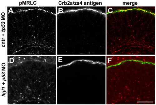
pMRLC localization in retinal neuroepithelia. (A,D) Immunofluorescence of phospho-Myosin regulatory light chain of Myosin II. (B,C) Immunofluorescence of Crumbs2a (Crb2a/zs4 antigen) to highlight the apical domain of retinal neuroepithelia. (C,F) Colocalization of pMRLC and zs4. Embryos were injected with either 8 ng control + 8ng tp53 MO (upper) or 8 ng tp53 + 8ng llgl1 ATG MO (lower). Scale bar: 50 μm. |
Expression Data
| Antibodies: | |
|---|---|
| Fish: | |
| Knockdown Reagents: | |
| Anatomical Terms: | |
| Stage: | Prim-25 |
Expression Detail
Antibody Labeling
Phenotype Data
Phenotype Detail
Acknowledgments
This image is the copyrighted work of the attributed author or publisher, and
ZFIN has permission only to display this image to its users.
Additional permissions should be obtained from the applicable author or publisher of the image.
Full text @ Development