
Fig. 4
- ID
- ZDB-FIG-120720-58
- Publication
- Okuda et al., 2012 - lyve1 expression reveals novel lymphatic vessels and new mechanisms for lymphatic vessel development in zebrafish
- Other Figures
- All Figure Page
- Back to All Figure Page
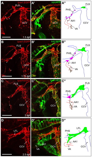
Distinct lymphangioblast populations contribute to the development of the LFL. (A-D′′) Lateral images of lyve1:EGFP expression (A-D) and both lyve1:EGFP and kdrl:RFP expression (A′-D′) of the lyve1:EGFP;kdrl:RFP transgenic at 1.5 dpf (A), 1.75 dpf (B), 2 dpf (C) and 2.5 dpf (D), with schematic diagrams of arteries (red), veins (blue) and lymphatic (green) at each stage (A′′-D′′). Lymphangioblast populations are indicated by asterisks. The PHS-L is in pink; the VA-L is in grey. AA1, mandibular arch; CCV, common cardinal vein; FLS, facial lymphatic sprout; LFL, lateral facial lymphatic; PHS, primary head sinus; PHS-L, primary head sinus lymphangioblast; VA, ventral aorta; VA-L, ventral aorta lymphangioblast. Scale bars: 100 μm. |